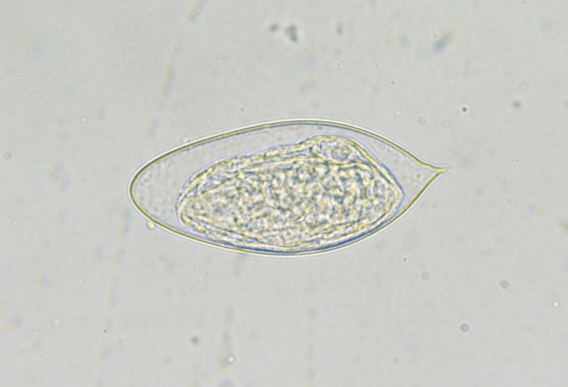
Examine the eggs of Schistosoma hematobium, noting the terminal spine, which acts to lodge the eggs in the blood vessels surrounding the urinary bladder. Eggs of S. mansoni have a lateral spine, and eggs of S. japonicum do not possess a spine. Attempt to view the seam of the operculum.
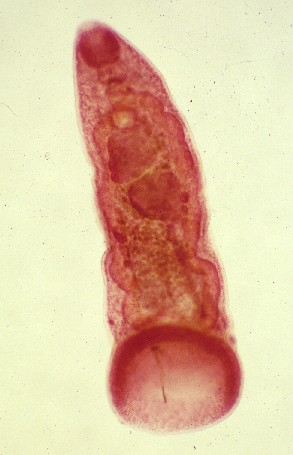
Examine the miracidium of Fasciola hepatica, noting the apical organ, pigmented photoreceptor, and ciliated outer layer. Compare to the electron micrograph on page 110 of your text (Fig. 4.11).
Examine the slide containing rediae and cercariae, comparing the rediae to Fig. 4.13 in your textbook (page 111). Note the mouth and “stomach”. Keep the slide on the microscope to examine the cercariae.
Examine the metacercariae of Diplostomum huronense and Posthodiplostomum minimum, and compare to Figure 2.1. Diplostomum huronense occurs free in the eye of fishes, and P. minimum encysts in the liver, on the heart, in the ovaries, and on the mesentery of fishes. Both species mature in bird definitive hosts, primarily by extension of the hindbody. Note the presence of Brande’s organ, which is an accessory organ for attachment found only in the metacercaria stage.
Cercariae with 2 eyespots, encysting on vegetation, Adults with ventral sucker at posterior end.
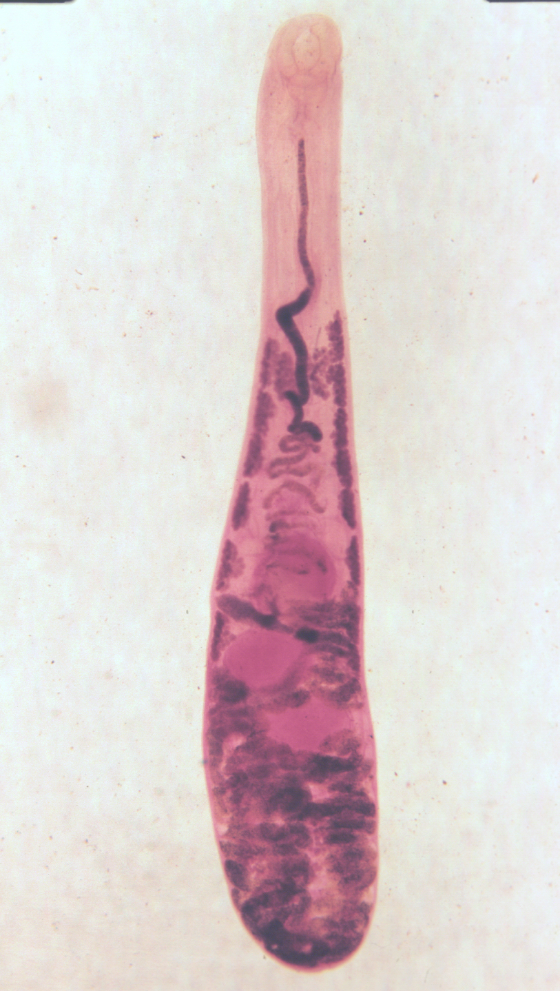
Megalodiscus americanus
Adult Megalodiscus spp. inhabit the cloacal region of frog intestines. Use Fig. 4.9 of
your text (page 108) as a guide to examining the anatomy of this specimen. Note the
large, posteriorly located, ventral sucker, and post-testicular ovaries.
Large cercariae lacking eyespots, and having strong tails. Cercariae develop in rediae and encyst on vegetation. Some members (Family Echinostomatidae) with a “spined collar” on cercariae, metacercariae, and adults. ventral sucker always midventral.
Fasciola hepatica (sheep liver fluke)
Use Fig. 4.9 of your text (page 108) as a guide to examining the anatomy of this fluke. Note the dendritic (branched), tandem testes in the posteror region, and the dendritic ovary, just posterior to the ventral sucker. Note also the outpocketings of the ceca.
Fasciolopsis buski (human intestinal fluke)
The anatomy of this species is very similar to F. hepatica, but the shape of the ventral sucker is different, and the ceca do not have outpocketings.
Furcocercous cercariae with eyespots that penetrate intermediate or definitive host. No redia stage.
Schistosoma mansoni
Adults in mesenteric veins of humans. Examine the slide of the adult male and female
in copula, with the female enclosed within the gynecophoral canal. The female anatomy
likely will be obscured by the male, but note the numerous testes in the male. Note
also that the ceca reunite in the male, forming a cyclocoel.
Gymnocephalous, lophocercous or pleurolophocercous opthalmocercariae. redia stage present.
Clonorchis sinensis (Chinese liver fluke)
Adults in bile ducts of humans. Note the lobed overy and the dendtitic, tandem testes.
The genital pore is located just anterior to the ventral sucker. There is no cirrus sac
or pars prostatica present.
Lophocercous, microcercous, or cotylocercous xiphidiocercariae. Redia absent.
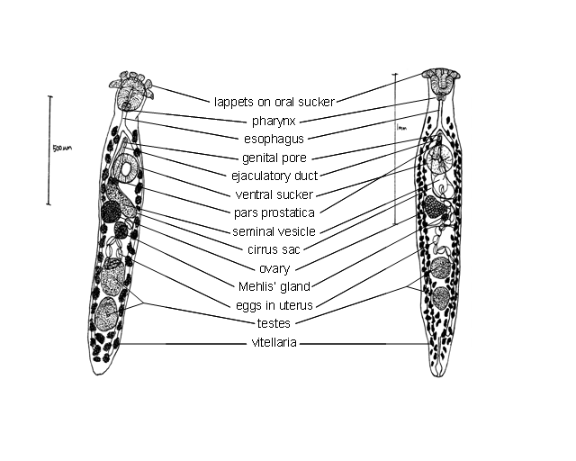
Crepidostomum sp.
Adults in small intestine of freshwater fishes. Examine your specimen of
Crepidostomum cornutum or
C. cooperi, and compare it to the appropriate
species on Figure 2.2.
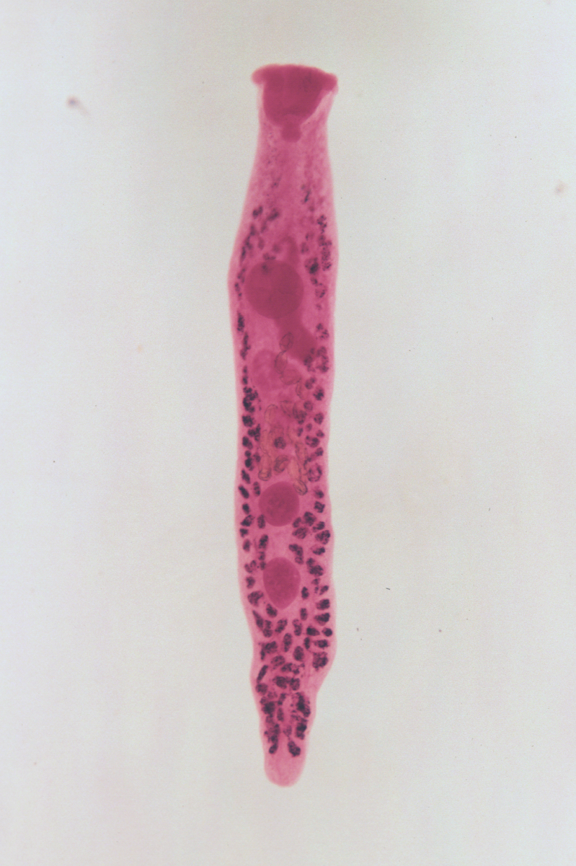
Prosthogomimus macrorchis
Adults in oviducts of birds. Note the opposite and entire testes, lobed ovary, and
the anterior location of the genital pore.
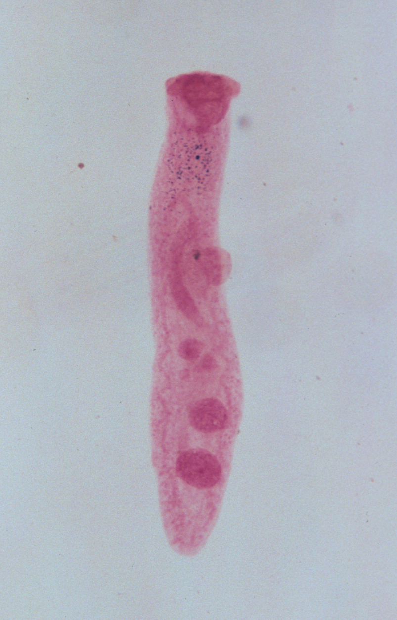
Hematoloechus meridionalis
Adults in lungs of rainforest frogs (Rana vaillanti). Note the reduced ventral
sucker, extensive uterus, and the large seminal receptacle near the ovary.